Description
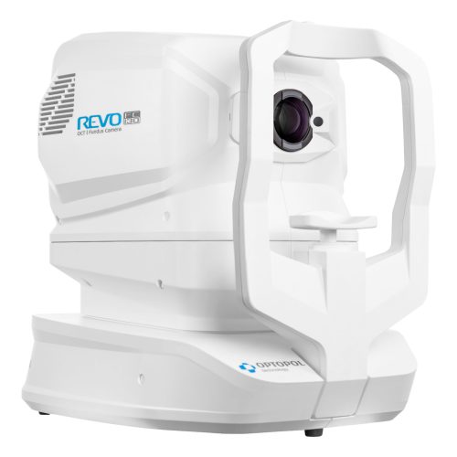
Spectral-Domain OCT | Color Fundus Camera
OCT made simple.
The REVO FC combines the world’s fastest SD-OCT with a non-mydriatic color fundus camera. Featuring our all-new AccuTrack™ real-time hardware-based eye tracker. Operating is as simple as the push of a button.
Technical Specifications
FUNDUS CAMERA |
|
| Type | Non-mydriatic fundus camera |
| Photography type | Color |
| Angle of view | 45° ± 5% |
| Min. pupil size for fundus | 3.3 mm |
| Camera | 12.3 Megapixel |
| Photography | Fundus (Retina, Central, Disc, Manual fixation), Anterior photo |
| Flash adjustment, Gain, Exposure | Auto, Manual |
| Intensity levels | High, Normal, Low |
OPTICAL COHERENCE TOMOGRAPHY |
|
| Technology | Spectral Domain OCT |
| Light Source | SLED Wavelength 850 nm |
| Bandwidth | 50 nm half bandwidth |
| Scanning speed | 80 000 measurements per second |
| Axial resolution | 2.8 μm digital, 5 μm in tissue |
| Transverse Resolution | 12 μm, typical 18 μm |
| Overall scan depth | 2.8 mm / ~6 mm in Full Range mode |
| Min. pupil size for OCT | 2.4 mm |
| Focus adjustment range | -25 D to +25 D |
| Scan range | Posterior 5 mm to 15 mm, Angio 3 mm to 9 mm, Anterior 3 mm to 18 mm |
| Scan types | 3D, Angio¹, Full Range Radial, Full Range B-scan, Radial (HD), B-scan (HD), Raster (HD), Raster 21 (HD), Cross (HD), TOPO ¹, Biometry AL¹ |
| Fundus alignment | IR, Live Fundus Reconstruction |
| Alignment method | Fully automatic, Automatic, Manual |
| Fundus Tracking | Real time active, iTracking |
| Retina analysis | Retina thickness, Inner Retinal thickness, Outer Retinal thickness, RNFL+GCL+IPL thickness, GCL+IPL thickness, RNFL thickness, RPE deformation, MZ/EZ-RPE thickness |
| Angiography OCT¹ | Vitreous, Retina, Choroid, Superficial Plexus, RPCP, Deep Plexus, Outer Retina, Choriocapilaries, Depth Coded, SVC, DVC, ICP, DCP, Custom, Enface, FAZ, VFA, NFA, Quantifi cation: Vessel Area Density, Skeleton Area Density, Thickness map |
| Glaucoma analysis | RNFL, ONH morphology, DDLS, OU and Hemisphere asymmetry, Ganglion analysis as RNFL+GCL+IP and GCL+IPL, Structure + Function ³ |
| Angiography mosaic | Acquistion method: Auto, Manual Mosaic modes: 10 mm x 6 mm, Manual up to 12 images |
| Biometry OCT ¹ | AL, CCT, ACD, LT, P, WTW |
| IOL Calculator ² | IOL Formulas: Hoffer Q, Holladay I, Haigis, Theoretical T, Regression II |
| Corneal Topography Map ¹ | Axial [Anterior, Posterior], Refractive Power [Kerato, Anterior, Posterior, Total], Net Map, Axial True Net, Equivalent Keratometer, Elevation [Anterior, Posterior], Height, KPI (Keratoconus Prediction Index) |
| Anterior (no lens/adapter required) |
Anterior Chamber Radial, Anterior Chamber B-scan, Pachymetry, Epithelium map, Stroma map, Angle Assessment, AIOP, AOD 500/750, TISA 500/750, Angle to Angle view |
| Connectivity | DICOM Storage SCU, DICOM MWL SCU, CMDL, Networking |
| Fixation target | OLED display (the target shape and position can be changed), External fixation arm |
| Dimensions (LxWxH) / Weight | 479 mm × 367 mm × 493 mm / 30 kg |
| Power supply / consumption | 100 V to 240 V, 50/60 Hz / 90 VA to 110 VA |
¹ An optional software module
² The Biometry module and a separate license for the IOL Calculator are required
³ Via connection with PTS software version 3.4 or higher